 Fraunhofer Institute for Microstructure of Materials and Systems IMWS
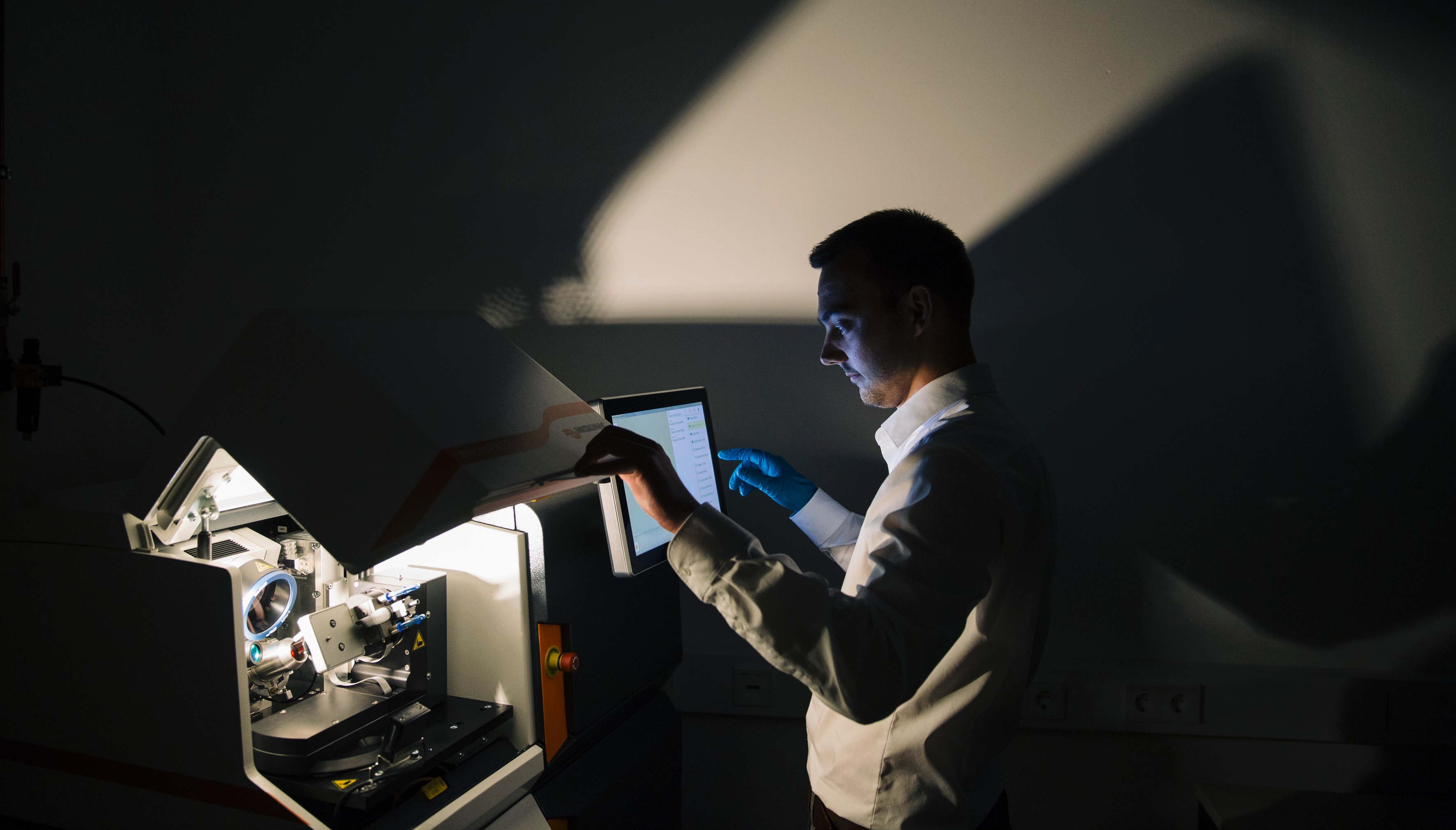
Georg Schusser works on the easy operability of microPREP, a device for laser-based sample preparation.

Each analysis is only as good as the quality of the specimen. This also applies to microstructure analyses of materials. Together with 3D-Micromac AG, we at the Fraunhofer IMWS have developed the microPREP system: State-of-the-art laser technology can be used to prepare ultrathin material specimens for electron microscope examination.
Dr. Thomas Höche carefully fixes the specimen of a semiconductor in the holder of the microPREP™ device, folds down the cover and starts the program. While a laser beam prepares the sample inside the device, Höche answers the questions of the potential buyers at one of the most important conferences for electron microscopy, the Microscopy Conference in Göttingen. Dr Höche, head of the development of microPREP™ at the Fraunhofer IMWS, particularly emphasizes the precision and reproducibility of the method. Only 15 minutes after starting the process he presents the finished, prepared specimen. The experts, responsible in their companies for material development and quality control, are impressed. With conventional methods they would need up to a whole day for this step.
Dr. Höche heads the nanomaterials and nanoanalysis group in the Fraunhofer IMWS; he is also an extraordinary Professor of Experimental Physics at the University of Leipzig. The experimental physicist came to Halle in 2010 with industrial experience. And thus at the same time returned to his roots. When he looks out of the window of his office he can see the Max Planck Institute for Microstructure Physics. »Over there is where I wrote my diploma thesis in 1991«, he says. At that time it was where the Institute of Solid State Physics and Electron Microscopy of the GDR’s Academy of Sciences was accommodated.
Following German reunification, a large part of the former Academy Institute became a Max Planck Institute; two workgroups were incorporated into the Fraunhofer IWM in Freiburg. As Höche returned to Halle, after time spent in Stuttgart, Jena, Berlin, Leipzig and Chemnitz, the two former workgroups of the Academy Institute had developed into a flourishing research facility, which initially belonged to the Fraunhofer IWM Freiburg and since 2016 has operated as an independent institute – the Fraunhofer IMWS.
Just how important good preparation methods are in microstructure analysis is experienced by Höche daily. Because one of the main pillars of the Fraunhofer IMWS is electron microscope examination of all kinds of materials – from semiconductors from the automotive and entertainment electronics industries through to oxide functional materials. To this end, the Fraunhofer IMWS has several transmission electron microscopes (TEM), including the TITAN, Fraunhofer's only aberration-corrected TEM. TITAN can be used to make the crystal structure, chemical element distribution and individual atoms in material specimens visible. In principle it works like a light microscope except that instead of light, electrons penetrate the specimen and produce imageable signals for further analysis.
In order for a specimen to be electron transparent, it must not be thicker than several tens of nanometres – a gigantic challenge for specimen preparation. Mechanical grinding damages the surface by forming defects. Preparation using focussed ion beams causes less damage, but is extremely time-consuming due to its low removal rate. Until now it has been feared that if lasers are used the material is heated and changes its structure.
Höche had the idea of using ultra-short pulsed lasers for the »athermal« specimen preparation back in 2010. At the Fraunhofer IMWS he continued his idea with Dr. Michael Krause. »We use a picosecond laser with pulse lengths of one billionth of a second, to avoid heating the specimen material«, explained Krause. Krause and Höche developed the microPREP™ system together with 3D-Micromac AG in Chemnitz – Höche’s former employer. In the prototype the specimen preparation takes place in two steps: A piece of the specimen material cut to size mechanically is given a defined shape with the ultra-short pulse laser and at the same time can be labelled – which excludes mix-ups and is very much approved of by our clients. In the second step, in a certain area of the specimen, an area is >>thinned<< to less than 15 micrometres, here the laser works parallel with the surface. Following the laser preparation with microPREP™, the specimen is only briefly polished in a wide ion beam system or a focussed ion beam before it can be examined in the TEM. The microPREP™ developers are proud to offer specimen preparation that is not only faster, but is also much less expensive.
Michael Krause is currently creating »recipes« for the preparation of all kinds of different materials. He is working on this with Georg Schusser. The laser engineer came to the Fraunhofer IMWS Halle from 3D-Micromac six months ago. Their aim is to make operating microPREP™ as easy as possible. To do this they are using, above all, the touchscreen on which it is possible to zoom and navigate in the same way as on a smartphone. With the help of the built-in camera it is even possible to mark on the screen the precise part of the specimen to be removed.
Following a successful presentation of the system at the Microscopy Conference in Göttingen, the first device has now been delivered to a customer in Asia.
Project manager Höche would like to develop microPREP™ into platform technology for the preparation of all kinds of different samples for different analysis methods. In particular, he has X-ray microscopy in his sights. »This method requires cylindrical samples with around 50 micrometre diameter«, explained the physicist. »In our device we only need to change the sample holder to be able to prepare these cylinders.« He is already sure to receive the attention of the experts at the next trade fair. Because X-ray microscopy is becoming increasingly important in microstructure analysis.
Text: Christine Broll
Fraunhofer IMWS and 3D-Micromac win TÜV SÜD Innovation Award 2018