Recovering more quickly after a fall: an app designed to improve patient care following pelvic ring fractures


Visual impairment, the abandonment of daily routines because of reduced mobility or occasional bouts of dizziness increase the chance of falls among elderly people. Around one half of such falls results in injury in which the pelvic ring is often affected. In a joint project with BG Klinikum Bergmannstrost Halle (gGmbH), the Fraunhofer Institute for Microstructure of Materials and Systems IMWS is developing a model designed to improve both post-operational care and rehabilitation following pelvic ring fractures. The resulting improvement in the quality of patient care will help those affected to regain their mobility more quickly while at the same time reducing nursing effort and expenses.
Elderly people who are already past retirement age are predominantly affected by pelvic ring fractures. This is attributable both to a sharp increase in the likelihood of falls in older people and to the deteriorating quality of their bones. When aggravated by bone atrophy (osteoporosis) or reductions in bone mass (osteopenia), a fall from a standing position is sufficient to cause damage to bones in the pelvic ring (fragility fractures). Apart from pain, patients may also be confined to bed, which can considerably increase the amount and cost of nursing care and mortality.
In the case of severe osteoporosis, the pelvis may even fracture if it is subject to normal stresses – not involving a fall. These types of fractures, insufficiency fractures, are related to demographic changes and predominantly affect women. They are caused by cavities inside the pelvis (‘alar voids’), which are characterized by significantly lower bone masses thus rendering the entire pelvis instable when subject to stress.
“Up to now there has been no consensus as to how best to surgically treat age-specific pelvic fractures. At the same time our society is ageing rapidly and the number of persons affected has been rising for years. We want to decisively improve the surgical treatment, therapeutic methods and nursing care of osteoporotic pelvis fractures and set clinical standards”, explains Dr. Thomas Mendel, trauma surgeon and head of the project at the Bergmannstrost hospital. Dr. Stefan Schwan, who coordinates the project at the Fraunhofer IMWS, adds: “There is a lack of knowledge about the effects of individual surgical methods or implants and precise insights on the mechanical behavior of a pelvic ring affected by osteoporosis and how it interacts with ligament, muscle and bone properties in older people.” It is precisely this interplay that the project partners are keen to reproduce in a virtual model.
“Simple numerical models for this problem already exist. However, such models neither take into account the marked microstructural changes in the bone and the associated high degree of alteration of mechanical properties nor the changes in the ligamentous apparatus responsible for channeling the stresses in and out of the pelvic ring system. For example, a tissue matrix can be found at those places where osteoporosis leads to a reduction in bone density whose characteristics for the failure behavior of the pelvic ring are important. Similarly, the role of the surrounding ligamentous apparatus, joints and muscles are currently not contemplated or only minimally contemplated in models”, explains Schwan.
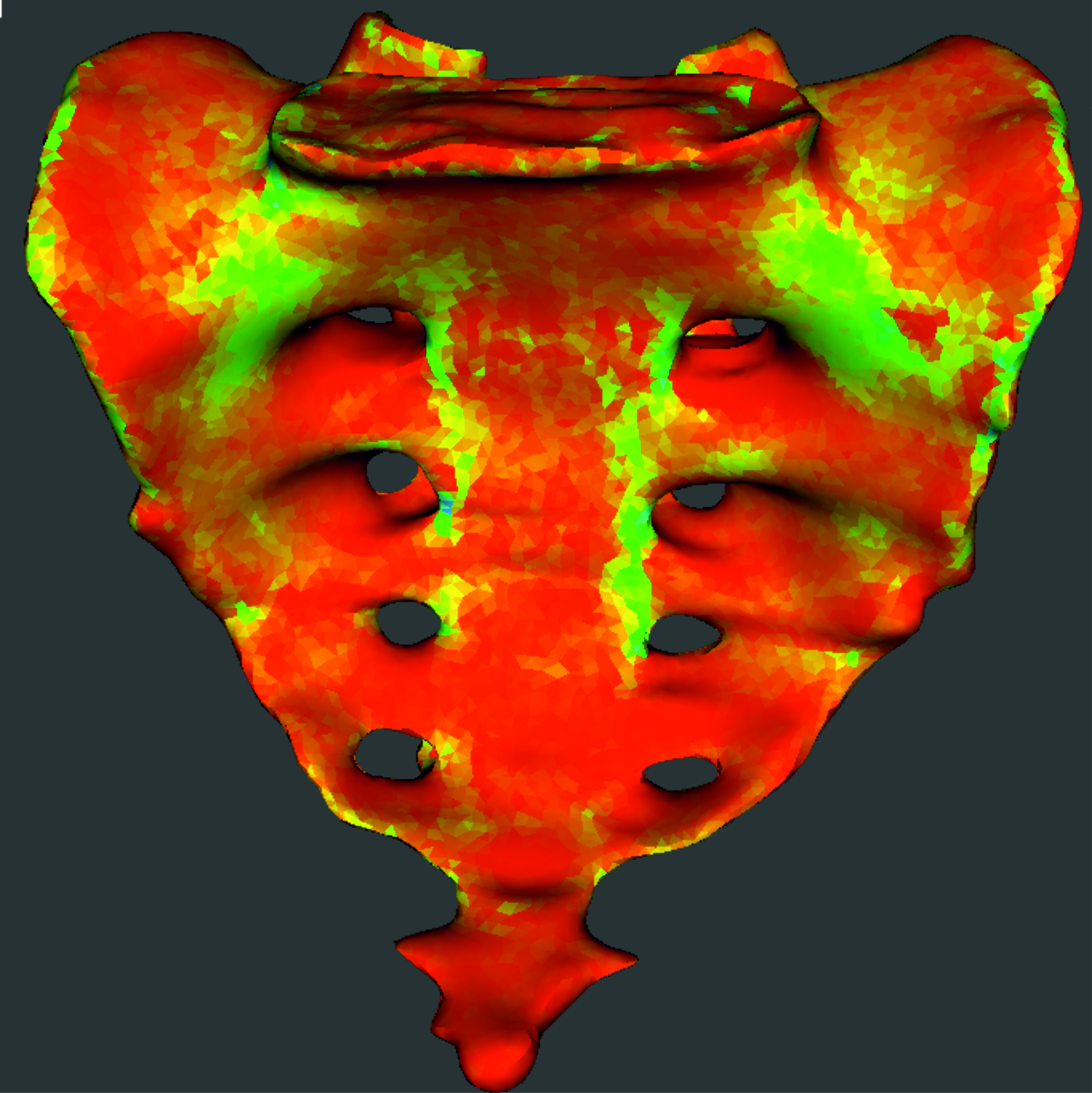
This is why the project involves the use of the finite element method (FEM), which enables a complete, three-dimensional representation of bone properties and stress fields in bone structures. Radiological data from magnetic resonance tomography (MRT), computer tomography and micro-computer tomography (µCT) all flow into this multiphase model, so that the geometry of the bone, its internal structure and the configuration of the surrounding soft tissue are recorded. The research team, which also includes rehab physicians and physiotherapists, links the datasets thus obtained with experimental micromechanical material models at the Fraunhofer IMWS. With the aid of algorithms which will be developed during the project, the model will then be able to specify material properties (elasticity modulus, thickness). This will allow for example the stresses exerted on the pelvic ring while walking to be realistically simulated and the origin and spread of fractures to be understood.
The resulting model will be made available in an app, which based on the data will provide information on suitable surgical techniques for optimal fracture treatment, suitable implants and tailored rehabilitation strategies. “If we are successful we will be able to offer an innovative and integral treatment method of the highest quality, from the moment the accident occurs to rehabilitation”, says Schwan summarizing the goals. “This will also help maintain the autonomy of elderly people and improve their medium-range life expectancy following a pelvic ring fracture.”